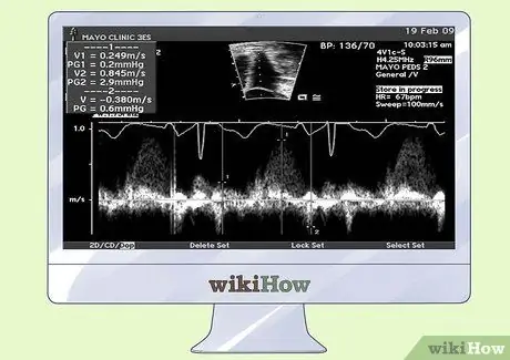
An echocardiogram is a non-invasive diagnostic test that examines the heart for abnormalities in the morphology and function of the heart chambers, valves, and myocardium. The test uses ultrasound waves to create a dynamic image of the heart in action. Echocardiograms are usually performed by a technician and the results of the echocardiogram are read by cardiologists. If you want to know how to interpret echocardiograms, you can familiarize yourself with some of the basics of the test. However, it is important that there is a doctor trained to analyze the echocardiogram to ensure an accurate diagnosis.
Steps

Step 1. Analyze the results of the echocardiogram to check for irregularities in the size and movement of the heart
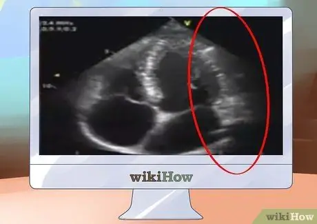
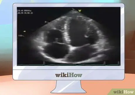
The dimensions are taken to ensure that the heart has not enlarged, which would indicate organ fatigue. If you have had echocardiograms previously, you can compare the echocardiogram results of each test to also determine if there have been any changes in the overall size of the heart.

Step 2. Measure the strength of your heart's ability to pump blood through the chambers
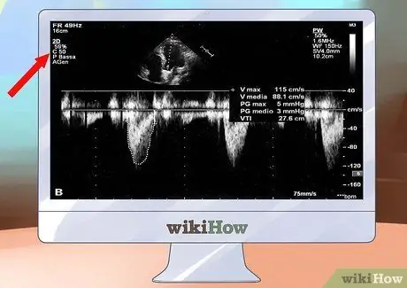
The pumping action is typically described as "ejection fraction" and should be between 55 and 65 percent. A lower ejection fraction can indicate systolic heart failure, while a higher percentage could mean diastolic heart failure. This test can also determine the reason for the abnormal reading, such as an area of the heart that has been weakened by a heart attack or a genetic condition that could increase the risk of heart problems in the future.

Step 3. Evaluate the thickness of the heart muscle wall with the echocardiogram results
A thickened wall around the heart means that the heart is not able to release and fill with blood as much. An elongated heart wall indicates that there may be weakening of the heart, due to a medical condition. When the muscle is the right size and functioning properly, the heart is able to easily fill with enough blood to meet all of the body's needs and then pump the blood out again.

Step 4. Examine the heart's four valves to determine if each is functioning properly
When doctors learn to interpret echocardiograms, they need to check the valves to make sure that blood is flowing properly through the heart. Leaking or improperly closing valves can slow blood flow and put the heart under excessive strain. A leaking valve can be detected if blood is seen flowing back through the heart. This problem may need to be treated with drugs or surgery, but the original diagnosis is typically made through the results of the echocardiogram.

Step 5. Determine the volume of blood that is circulating through the heart
This echocardiogram result could be directly influenced by drugs such as diuretics. A low volume may indicate that the heart is not pumping blood through the body as efficiently as it should. This problem could be caused by a number of conditions affecting the heart and cardiovascular system.


