A microscope is a device that magnifies an image allowing you to see small structures in detail. Although there are several models of various sizes, studio and home models typically have similar components: a base, an eyepiece, a lens, and a storage table. By learning the basic techniques of using a microscope, you can protect it from damage and have a valuable study tool.
Steps
Part 1 of 3: Assemble the Microscope
Step 1. Know the various components
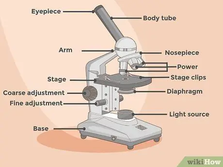
There are several essential pieces that you need to know how to recognize and use correctly. The eyepiece is the element that allows you to look through the microscope and observe the sample; the simple models are equipped with only one eyepiece, while the more complex ones can be binocular. Here are the components:
- The stage is the platform on which the slides to be observed are placed.
- The stand is the structure that connects the base to the eyepiece.
- There are two focus knobs: the fine adjustment knob (micrometric screw) and the coarse adjustment knob (coarse screw). The second has usually larger dimensions, is located on the side of the microscope and allows you to move the objective, moving it away or closer to the sample; allows you to view the sample and coarsely focus its image. The micrometer screw is smaller and is used to view details; it is used to precisely focus on what you are observing.
- The lens is the element that magnifies the image; there are different types for different magnification levels.
- The light source (lamp) is located at the base of the instrument and is directed towards the storage table; provides the lighting needed to view the image.
- The condenser is located just below the stage and allows you to vary the amount of light that hits the sample.
Step 2. Place the microscope on a clean, flat surface
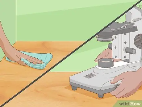
Eliminates all residues that could potentially damage the instrument; clean the table with an all-purpose cleaner and a microfiber rag if needed. Make sure there is a power outlet nearby.
- Carry the microscope holding it by the base and the stand; never lift it by grabbing it only by the stand.
- Put it on the table and insert the plug into the electrical outlet.
Step 3. Have the instrument manual available
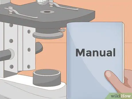
Read it carefully, if you want to have specific instructions for handling the model in your possession; the manual should also include instructions on maintenance and cleaning.
- Keep the manual close to the microscope for easy reference.
- If you have lost it, search online to find a downloadable copy from the microscope manufacturer's website; if you can't find it online, contact the manufacturer and ask for a copy to be sent to you.
Part 2 of 3: Prepare the Slides
Step 1. Wash your hands before starting
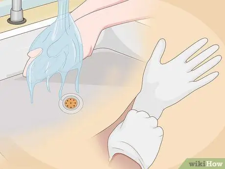
The hands are covered with sebum which can be easily transferred to the slides and samples. The sebum damages both the instrument and the slides; if you have gloves on hand, they are worth putting on.
Keep your hands and work area free of dust and contaminants as much as possible
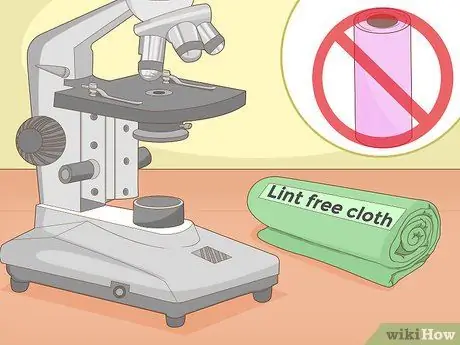
Step 2. Have a microfiber cloth handy to clean and touch the slides
It is a special fabric that does not leave lint or other fibers on the surface it comes into contact with. Many slides have an electrical charge on one side to simplify specimen fixing operations; however, this feature makes them susceptible to attracting dust and other debris. A lint-free cloth helps counter contamination.
- Never use paper towels to clean slides, as they leave a lot of fibers and lint.
- If you are wearing gloves, you can touch the slide, but try to grab it only by the edges.
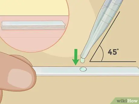
Step 3. To get started, use prepared slides
They already contain an appropriately fixed sample; you can buy them in stores that sell scientific material and they are often included in the microscope package. Once you are familiar with the tool, you can try to prepare the slides yourself.
- First, you need to get the sample you want to observe in detail; pond water or pollen are perfect things to start with.
- Drop a small drop of water or place a few pollen spores directly on the slide.
- Place a coverslip 45 ° over the first and drop it on the sample; the water should hold it in place.
- To keep the samples longer, add some nail polish along the edges of the slide to secure that coverslip.
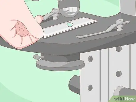
Step 4. Place the slide on the stage
Lift it by grabbing it at the edges to avoid leaving fingerprints; remember that sebum and fingerprints can contaminate it. You can also use the microfiber cloth to lift the slide.
If it is dirty, wipe it gently with the cloth
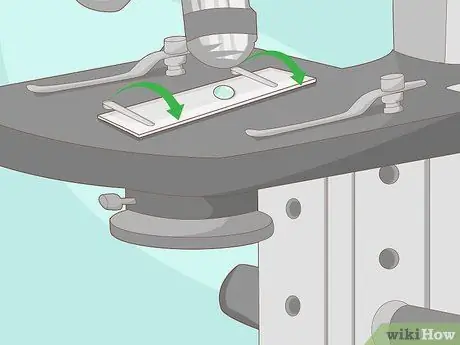
Step 5. Secure the slide to the stage using the two clips
On the table there are two clips (plastic or metal) that are used to fix the slide in place, allowing you to have your hands free and focus the image; you should be able to insert the slide under the clips without difficulty.
- Do not force the slide under the clips, which should rise slightly to aid insertion. If you have difficulty, try to fit it under one clothespin at a time; lift the clip, slide the slide under it and then repeat with the second.
- Remember that slides are quite fragile and can break if you don't handle them properly.
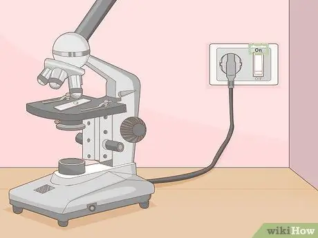
Step 6. Turn on the microscope
The switch is usually located on one side. The center of the slide should glow with a disc of light.
- If you don't see any light, try adjusting the condenser until it's fully open. This element should be equipped with a lever or a rotating disk, which manages its diameter and modifies the amount of light that can filter; if the condenser is closed, you don't see any light. Move the lever or disc until the light beam appears.
- If that doesn't fix the problem, check the electrical outlet or call for service to change the microscope bulb.
Part 3 of 3: Focus with the Microscope
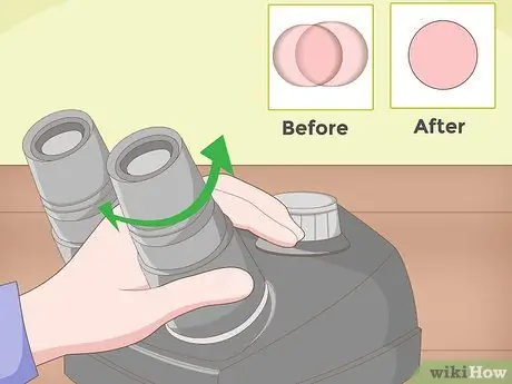
Step 1. Adjust the eyepieces if it is a binocular model
If your microscope is a monocular type, you can skip this step. Rotate each eyepiece to find the correct distance between the eyes, ie the interpupillary distance; when you look through both elements, you should see only one disc of light.
- If you see two images, you need to continue adjusting the distance between the eyepieces.
- Push them together or distance them until you see a single bright disc.
- Take off your glasses, if you wear them; you can use the tool's focus settings to fit the view.
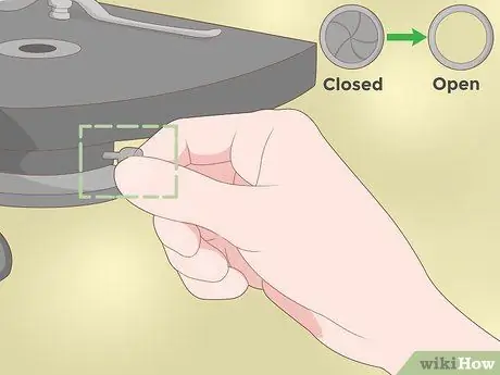
Step 2. Adjust the capacitor to the maximum amplitude
This element allows you to change the amount of light hitting the slide; to start focusing on the specimen, you need to brighten it to the max. There should be a lever or wheel that allows you to change the diameter of the opening.
Move the lever or wheel until the condenser is fully open
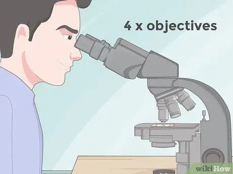
Step 3. Start focusing the image using the lower magnification power
Probably, the microscope has two or three objectives mounted on a rotating disk, which you can rotate to change magnification. You should start with the 4x power one and increase the value until the image is in focus; typically, the 4x (or 3.5x) objective is the minimum standard for a basic microscope.
- The low-magnification lens allows for a larger field of view and allows you to focus the image slowly, without losing reference points. In English, this objective is called "scanning", precisely because it allows you to gradually study the sample; starting with a higher magnification you may not see the sample in its entirety or you may miss important reference points.
- The two most powerful commonly used lenses are 10x and 40x.
- The eyepiece has a magnification power of 10x which is multiplied by that of the objective in use; as a result, a 4x objective provides a total magnification of 40x, a 10x objective provides a 100x magnified image, and the 40x objective yields a total magnification of 400x.
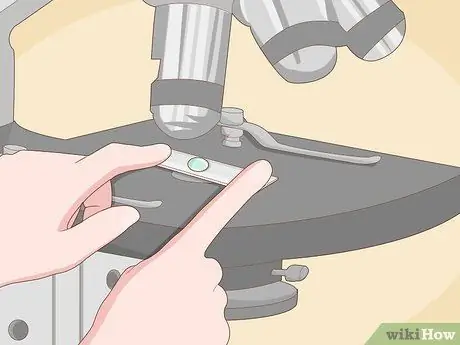
Step 4. Move the slide to center it on the stage if necessary
Most slides are considerably larger than the specimen attached to them. If you want to observe a substance, try to place it directly in the center of the light source; if you can't see it, slowly move the slide while looking into the eyepiece.
Remember that magnification offers an upside-down image, so you have to move the slide in the opposite direction to view it correctly through the lenses
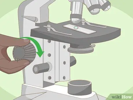
Step 5. Focus the image using the adjustment screws and the condenser
Start by operating the coarse screw (the larger one), then move on to the micrometric screw for fine adjustments and finally change the lighting level. As you look through the eyepiece, slowly turn the coarse screw until the image begins to focus.
- Use the micrometer screw to optimize the slide image.
- Be aware that as you focus the image, the stage will rise as it approaches the objective and it is possible for it to reach a level where the slide touches some of the objective lenses; so be very careful during the procedure, to prevent this from happening.
- Adjust the condenser located under the stage. By reducing the amount of light, you can get a sharper image with better contrast.
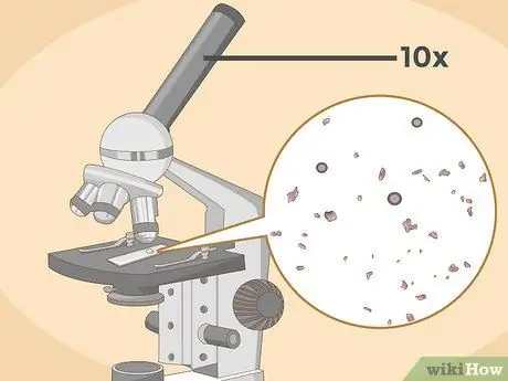
Step 6. Enlarge the image using the strongest lens
Move to a higher level only when you are unable to focus further on the details with the less powerful lens; a higher magnification allows you to see more details of the sample. Not all objectives are used to observe all slides, as some focus too closely.
- Be careful when changing lenses to avoid breaking the slide.
- Use the micrometer screw when using a high magnification, for example 10x, because the coarse screw moves the objective too close to the stage, with the risk of breaking the slide.
- Switch between lenses and adjust the focus screws until you are familiar with the tool; try to use different slides to improve skills.
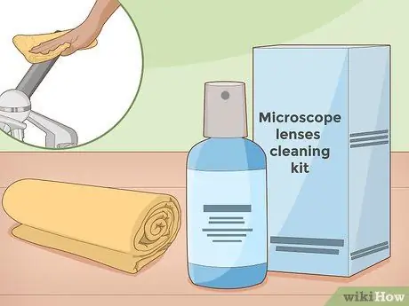
Step 7. Place the microscope in a dust bag
Lenses are easily damaged by dust and other particles suspended in the air; by keeping the lenses and the stage clean, you prevent this type of damage. Clean the lenses only with a specific liquid and cloth.


